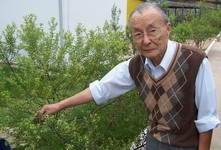
Prof. Yong Kun Park, M.D. Ph.D.
Médico patologista, bioquímico
professor da Faculdad de Ingeniería
de alimentos de UNICAMP.


Investigador visitante en diversos centros de investigación del mundo como; Rochester University, North Carolina University, Centro de control de enfermedades de las fuerzas armadas americanas, etc. Su principal interes es el uso de propoleos en biotecnologia e farmacologia.

Items 1 - 14 of 14

.

1: J Agric Food Chem. 2005 Dec 28;53(26):10306-9.

Related Articles, Links

Click here to read  
Suppressive effects of ethanolic extracts from propolis and its main botanical origin on dioxin toxicity.

Park YK, Fukuda I, Ashida H, Nishiumi S, Yoshida K, Daugsch A, Sato HH, Pastore GM.

Department of Food Science, College of Food Engineering, State University of Campinas, P.O. Box 6177, Campinas, SP, Brazil. ykpark@fea.unicamp.br

Suppressive effects of ethanolic extracts prepared from propolis group 12 and its main botanical origin (leaf bud of Baccharis dracunculifolia) on transformation of the aryl hydrocarbon receptor (AhR), the initial action of dioxin toxicity, were investigated. It was found that suppressive effects of propolis on AhR transformation were relatively higher than those of resins of its botanical origin in cell-free system and in Hepa-1c1c7 cells. When the composition of chemical ingredients was measured, propolis contained slightly higher amounts of flavonoid aglycones as compared with its botanical origin with the same characteristics. Moreover, antiradical activity, one of the typical biological activities of flavonoids, in propolis was also slightly higher than that in its botanical origin. These results indicate that not only propolis but also its botanical origin contains high amounts of flavonoid aglycones and that both of them are useful dietary sources for flavonoids with a potency to prevent dioxin toxicity.

PMID: 16366731 [PubMed - indexed for MEDLINE]


2: J Agric Food Chem. 2005 Feb 23;53(4):1166-72.

Related Articles, Links

Click here to read  
Some chemical composition and biological activity of northern Argentine propolis.

Isla MI, Paredes-Guzman JF, Nieva-Moreno MI, Koo H, Park YK.

Department of Food Science, College of Food Engineering, State University of Campinas, P.O. Box 6177, 13083-970, Campinas, Sao Paulo, Brazil.

Twenty-five samples of propolis were collected from seven different regions in northern Argentina; ethanolic extracts of propolis were prepared from all samples, and the respective samples were examined for UV absorption spectra, RPHPTLC, RPHPLC, antimicrobial activity, antiradical activity, and total phenolic content. It was found that 16 of the 25 samples showed a phenolic profile similar to that found in samples from southern Brazil and corresponding to poplar-based propolis and that the rest of the samples showed a different profile and higher antimicrobial and antiradical activities.

PMID: 15713035 [PubMed - indexed for MEDLINE]


3: Biosci Biotechnol Biochem. 2004 Apr;68(4):935-8.

Related Articles, Links

Click here to read  
Suppression of dioxin mediated aryl hydrocarbon receptor transformation by ethanolic extracts of propolis.

Park YK, Fukuda I, Ashida H, Nishiumi S, Guzman JP, Sato HH, Pastore GM.

State University of Campinas, College of Food Engineering (UNICAMP), Department of Food Science, Laboratory of Food Biochemistry, SP, Brazil. ykpark@fea.unicamp.br

Present study demonstrated that the ethanolic extracts of propolis containing higher concentrations of flavonoids suppressed 2,3,7,8-tetrachlorodibenzo-p-dioxin (TCDD)-induced aryl hydrocarbon receptor transformation in a dose-dependent manner. The IC(50) values of propolis group 3 and group 12 were 1.2 and 3.6 microg/ml, respectively, indicating that propolis showed stronger antagonistic effects as compared with vegetable extracts.

PMID: 15118327 [PubMed - indexed for MEDLINE]


4: J Agric Food Chem. 2004 Mar 10;52(5):1100-3.

Related Articles, Links

Click here to read  
Chemical constituents in Baccharis dracunculifolia as the main botanical origin of southeastern Brazilian propolis.

Park YK, Paredes-Guzman JF, Aguiar CL, Alencar SM, Fujiwara FY.

Department of Food Science, College of Food Engineering, State University of Campinas, P.O. Box 6177, 13083-970, Campinas, Sao Paulo, Brazil. ykpark@fea.unicamp.br

Previously, it was reported that one group of propolis (Group 12) was identified in southeastern Brazil, and the botanical origin of the propolis was Baccharis dracunculifolia resinous exudates. It was also observed that honeybee (Africanized Apis mellifera) mainly visited the leaf buds or unexpanded leaves of B. dracunculifolia but rarely expanded leaves. B. dracunculifolia is dioecious with male and female inflorescences, and RPHPLC of the ethanolic extracts of the respective male and female bud resinous exudates showed the same profiles. RPHPLC profiles of propolis G12 leaf buds and unexpanded and expanded leaves of B. dracunculifolia showed similarity, but unexpanded leaves quantitatively decreased in chemical constituents as compared with leaf buds. In the case of expanded leaves, all chemical constituents were severely decreased or disappeared. Artepillin C (3,5-diprenyl-4-hydroxycinnamic acid) was also identified in both propolis and resinous exudates, and both ethanolic extracts contained the highest concentrations of this compound as compared with the rest of the chemical constituents.

PMID: 14995105 [PubMed - indexed for MEDLINE]


5: Oral Microbiol Immunol. 2002 Dec;17(6):337-43.

Related Articles, Links

Click here to read  
Effects of apigenin and tt-farnesol on glucosyltransferase activity, biofilm viability and caries development in rats.

Koo H, Pearson SK, Scott-Anne K, Abranches J, Cury JA, Rosalen PL, Park YK, Marquis RE, Bowen WH.

Center for Oral Biology and Eastman Department of Dentistry, University of Rochester Medical Center, NY 14642, USA.

Propolis, a resinous hive product secreted by Apis mellifera bees, has been shown to reduce the incidence of dental caries in rats. Several compounds, mainly polyphenolics, have been identified in propolis. Apigenin and tt-farnesol demonstrated biological activity against mutans streptococci. We determined here their effects, alone or in combination, on glucosyltransferase activity, biofilm viability, and development of caries in rats. Sprague-Dawley rats were infected with Streptococcus sobrinus 6715 and treated topically twice daily as follows: (1) tt-farnesol, (2) apigenin, (3) vehicle control, (4) fluoride, (5) apigenin +tt-farnesol, and (6) chlorhexidine. Apigenin (1.33 mM) inhibited the activity of glucosyltransferases in solution (90-95%) and on the surface of saliva-coated hydroxyapatite beads (35-58%); it was devoid of antibacterial activity. tt-Farnesol (1.33 mM) showed modest antibacterial activity against biofilms and its effects on glucosyltransferases were minimal. The incidence of smooth-surface caries was significantly reduced by apigenin +tt-farnesol (60%), fluoride (70%), and chlorhexidine (72%) treatments compared to control (P < 0.05).

PMID: 12485324 [PubMed - indexed for MEDLINE]


6: Caries Res. 2002 Nov-Dec;36(6):445-8.

Related Articles, Links

Click here to read  
Effect of a mouthrinse containing selected propolis on 3-day dental plaque accumulation and polysaccharide formation.

Koo H, Cury JA, Rosalen PL, Ambrosano GM, Ikegaki M, Park YK.

Department of Dentistry, University of Rochester Medical Center, Rochester, NY, USA.

The aim of this study was to evaluate the effect of a mouthrinse containing propolis SNB-RS on 3-day dental plaque accumulation. Six volunteers took part in a double-blind crossover study performed in two phases of 3 days. During each phase the volunteers refrained from all oral hygiene and rinsed with 20% sucrose solution 5 times a day to enhance dental plaque formation and with mouthrinse (placebo or experimental) twice a day. On the 4th day, the plaque index (PI) of the volunteers was scored and the supragingival dental plaque was analyzed for insoluble polysaccharide (IP). The PI (SD) for the experimental group was 0.78 (0.17), significantly less than for the placebo group, 1.41 (0.14). The experimental mouthrinse reduced the IP concentration in dental plaque by 61.7% compared to placebo (p < 0.05). An experimental mouthrinse containing propolis SNB-RS was thus efficient in reducing supragingival plaque formation and IP formation under conditions of high plaque accumulation.
Copyright 2002 S. Karger AG, Basel

Publication Types:

·       Clinical Trial

·       Randomized Controlled Trial


PMID: 12459618 [PubMed - indexed for MEDLINE]


7: Antimicrob Agents Chemother. 2002 May;46(5):1302-9.

Related Articles, Links

Click here to read   Click here to read  
Effects of compounds found in propolis on Streptococcus mutans growth and on glucosyltransferase activity.

Koo H, Rosalen PL, Cury JA, Park YK, Bowen WH.

Center for Oral Biology and Eastman Department of Dentistry, University of Rochester Medical Center, Rochester, New York 14642, USA. Hyun_Koo@urmc.rochester.edu

Propolis, a resinous bee product, has been shown to inhibit the growth of oral microorganisms and the activity of bacterium-derived glucosyltransferases (GTFs). Several compounds, mainly polyphenolics, have been identified in this natural product. The present study evaluated the effects of distinct chemical groups found in propolis on the activity of GTF enzymes in solution and on the surface of saliva-coated hydroxyapatite (sHA) beads. Thirty compounds, including flavonoids, cinnamic acid derivatives, and terpenoids, were tested for the ability to inhibit GTFs B, C, and D from Streptococcus mutans and GTF from S. sanguinis (GTF Ss). Flavones and flavonols were potent inhibitors of GTF activity in solution; lesser effects were noted on insolubilized enzymes. Apigenin, a 4',5,7-trihydroxyflavone, was the most effective inhibitor of GTFs, both in solution (90.5 to 95% inhibition at a concentration of 135 microg/ml) and on the surface of sHA beads (30 to 60% at 135 microg/ml). Antibacterial activity was determined by using MICs, minimum bactericidal concentrations (MBCs), and time-kill studies. Flavanones and some dihydroflavonols, as well as the sesquiterpene tt-farnesol, inhibited the growth of S. mutans and S. sobrinus; tt-farnesol was the most effective antibacterial compound (MICs of 14 to 28 microg/ml and MBCs of 56 to 112 microg/ml). tt-Farnesol (56 to 112 microg/ml) produced a 3-log-fold reduction in the bacterial population after 4 h of incubation. Cinnamic acid derivatives had negligible biological activities. Several of the compounds identified in propolis inhibit GTF activities and bacterial growth. Apigenin is a novel and potent inhibitor of GTF activity, and tt-farnesol was found to be an effective antibacterial agent.

PMID: 11959560 [PubMed - indexed for MEDLINE]


8: J Agric Food Chem. 2002 Apr 24;50(9):2502-6.

Related Articles, Links

Click here to read  
Botanical origin and chemical composition of Brazilian propolis.

Park YK, Alencar SM, Aguiar CL.

Department of Food Science, College of Food Engineering, State University of Campinas, 13081-970 Campinas, Sao Paulo, Brazil. ykpark@fea.unicamp.br

Brazilian propolis has been classified into 12 groups based on physicochemical characteristics: five in the southern Brazil group (group 3), one in the southeastern Brazil group (group 12), and six in the northeastern Brazil group (group 6). The plant origins of these groups were investigated using reversed-phase high-performance thin-layer chromatography (RPHPTLC), reversed-phase high-performance liquid chromatography (RPHPLC), and gas chromatography-mass spectrometry (GC-MS). It was concluded that the origins of propolis group 3, group 6, and group 12 are resins of the poplar tree, Hyptis divaricata, and Baccharis dracunculifolia, respectively.

PMID: 11958612 [PubMed - indexed for MEDLINE]


9: J Nat Prod. 2001 Oct;64(10):1278-81.

Related Articles, Links

Click here to read  
Anti-AIDS agents.
48.(1) Anti-HIV activity of moronic acid derivatives and the new melliferone-related triterpenoid isolated from Brazilian propolis.

Ito J, Chang FR, Wang HK, Park YK, Ikegaki M, Kilgore N, Lee KH.

Natural Products Laboratory, School of Pharmacy, University of North Carolina, NC 27599, USA.

A new triterpenoid named melliferone (1), three known triterpenoids, moronic acid (2), anwuweizonic acid (3), and betulonic acid (4), and four known aromatic compounds (5-8) were isolated from Brazilian propolis and tested for anti-HIV activity in H9 lymphocytes. Moronic acid (2) showed significant anti-HIV activity (EC(50) <0.1 microg/mL, TI >186) and was modified to develop more potent anti-AIDS agents.

PMID: 11678650 [PubMed - indexed for MEDLINE]


10: Caries Res. 2000 Sep-Oct;34(5):418-26.

Related Articles, Links

Click here to read  
Effects of Apis mellifera propolis on the activities of streptococcal glucosyltransferases in solution and adsorbed onto saliva-coated hydroxyapatite.

Koo H, Vacca Smith AM, Bowen WH, Rosalen PL, Cury JA, Park YK.

Faculty of Dentistry of Piracicaba, State University of Campinas, Piracicaba, Brazil. hyunkoobr@yahoo.com

Propolis, a resinous hive product collected by Apis mellifera bees, has been used for thousands of years in folk medicine. Ethanolic extracts of propolis (EEP) have been shown to inhibit the activity of a mixture of crude glucosyltransferase (Gtf) enzymes in solution. These enzymes synthesize glucans from sucrose, which are important for the formation of pathogenic dental plaque. In the present study, the effects of propolis from two different regions of Brazil on the activity of separate, purified Gtf enzymes in solution and on the surface of saliva-coated hydroxyapatite (sHA) beads were evaluated. The EEP from Minas Gerais (MG; Southeastern Brazil) and Rio Grande do Sul (RS; Southern Brazil) were tested for their ability to inhibit the enzymes GtfB (synthesis of insoluble glucan), GtfC (insoluble/soluble glucan) and GtfD (soluble glucan). The effects of propolis on Gtf from Streptococcus sanguis (soluble glucan synthesis) was also explored. The EEP from both regions effectively inhibited the activity of all Gtfs in solution (75-95%) and on the surface of sHA beads (45-95%) at concentrations between 0.75 and 3.0 mg of propolis/ml. However, the two samples of propolis showed different levels of inhibition on each of the enzymes tested. In general, EEP RS demonstrated a significantly higher inhibitory activity on GtfB and C activities (both solution and surface assays) than EEP MG at concentrations between 0.047 and 0.187 mg/ml (p<0.05). EEP MG, on the other hand, exhibited a greater inhibitory effect on the activities of surface GtfD (at 0.375, 0.75 and 1.5 mg/ml) and S. sanguis Gtf (at 1.5 and 3.0 mg/ml; p<0.05). These data indicate that EEP is a potent inhibitor of Gtf enzymes in solution and adsorbed on an experimental pellicle; however, its effect on Gtf activity is variable depending on the geographical origin of the propolis samples. There is a need to identify the active compounds of propolis.

PMID: 11014909 [PubMed - indexed for MEDLINE]


11: Curr Microbiol. 2000 Sep;41(3):192-6.

Related Articles, Links

Click here to read  
Effect of a new variety of Apis mellifera propolis on mutans Streptococci.

Koo H, Rosalen PL, Cury JA, Ambrosano GM, Murata RM, Yatsuda R, Ikegaki M, Alencar SM, Park YK.

Department of Physiological Sciences, Faculty of Dentistry of Piracicaba, State University of Campinas, Caixa Postal 52, Piracicaba, 13414-900, SP, Brazil. Hyun-Koo@urmc.rochester.edu

The effects of a new variety of propolis, from Northeastern Brazil (BA), on growth of mutans streptococci, cell adherence, and water-insoluble glucan (WIG) synthesis were evaluated. Propolis from Southeastern (MG) and Southern (RS) Brazil were also tested as an extension of our previous work. Ethanolic extracts of propolis (EEP) were prepared and analyzed by reversed-phase HPLC. For the antibacterial activity assays, minimum inhibitory concentrations (MIC) and minimum bactericidal concentrations (MBC) of EEPs against Streptococcus mutans, S. sobrinus, and S. cricetus were determined. Cell adherence of S. mutans and S. sobrinus to a glass surface was measured spectrophotometrically at 550 nm. WIG synthesized from sucrose by glucosyltransferase (Gtf) was extracted and quantified by the phenol-sulfuric method. The HPLC profile of the new variety of propolis was entirely different from Southeastern and Southern propolis. Neither flavonoid aglycones nor p-coumaric acid were detected in EEP BA. All EEPs demonstrated biological activities against mutans streptococci; EEP BA showed the highest potency in all in vitro parameters evaluated in this study. The ranges of MIC values were 50 (EEP BA)-400 microg/ml (MG), for S. mutans; and 25 (BA)-400 microg/ml (MG), for S. sobrinus and S. cricetus. The bactericidal concentration of EEPs was four to eight times the MIC values. The adherence of S. mutans and S. sobrinus cells and WIG synthesis were markedly inhibited by EEPs, demonstrating significant inhibition at all concentrations compared with the control (80% ethanol) (p<0.05). EEP BA showed 80% inhibition of cell adherence and WIG synthesis at concentrations as low as 12.5 and 7.8 microg/ml, respectively. The results show that the new variety of propolis was exceptionally effective in all in vitro parameters tested against mutans streptococci; biological effects of propolis are likely not to be due solely to flavonoids and (hydroxy)cinnamic acid derivatives.

PMID: 10915206 [PubMed - indexed for MEDLINE]


12: Arch Oral Biol. 2000 Feb;45(2):141-8.

Related Articles, Links

Click here to read  
In vitro antimicrobial activity of propolis and Arnica montana against oral pathogens.

Koo H, Gomes BP, Rosalen PL, Ambrosano GM, Park YK, Cury JA.

School of Dentistry of Piracicaba, State University of Canipinas, Areiao, S.P., Brazil.

Arnica and propolis have been used for thousands of years in folk medicine for several purposes. They possess several biological activities such as anti-inflammatory, antifungal, antiviral and tissue regenerative, among others. Although the antibacterial activity of propolis has already been demonstrated, very few studies have been done on bacteria of clinical relevance in dentistry. Also, the antimicrobial activity of Arnica has not been extensively investigated. Therefore the aim here was to evaluate in vitro the antimicrobial activity, inhibition of adherence of mutans streptococci and inhibition of formation of water-insoluble glucan by Arnica and propolis extracts. Arnica montana (10%, w/v) and propolis (10%, w/v) extracts from Minas Gerais State were compared with controls. Fifteen microorganisms were used as follows: Candida albicans--NTCC 3736, F72; Staphylococcus aureus--ATCC 25923; Enterococcus faecalis--ATCC 29212; Streptococcus sobrinus 6715; Strep. sanguis--ATCC 10556; Strep. cricetus--HS-6; Strep. mutans--Ingbritt 1600; Strep. mutans--OMZ 175; Actinomyces naeslundii--ATCC 12104, W 1053; Act. viscosus OMZ 105; Porphyromonas gingivalis; Porph. endodontalis and Prevotella denticola (the last three were clinical isolates). Antimicrobial activity was determined by the agar diffusion method and the zones of growth inhibition were measured. To assess cell adherence to a glass surface, the organisms were grown for 18 h at 37 degrees C in test-tubes at a 30 degree angle. To assay water-insoluble glucan formation, a mixture of crude glucosyltransferase and 0.125 M sucrose was incubated for 18 h at 37 degrees C in test-tubes at a 30 degree angle. Arnica and propolis extracts (20 microl) were added to these tubes to evaluate the % of inhibition of cell adherence and water-insoluble glucan formation. The propolis extract significantly inhibited all the microorganisms tested (p < 0.05), showing the largest inhibitory zone for Actinomyces spp. The Arnica extract did not demonstrate significant antimicrobial activity. Cell adherence and water-insoluble glucan formation were almost completely inhibited by the propolis extract at a final concentration of 400 microg/ml and 500 microg/ml, respectively. The Arnica extract showed slight inhibition of the adherence of the growing cells (19% for Strep. mutans and 15% for Strep. sobrinus) and of water-insoluble glucan formation (29%) at these same concentrations. Thus, the propolis extract showed in vitro antibacterial activity, inhibition of cell adherence and inhibition of water-insoluble glucan formation, while the Arnica extract was only slightly active in those three conditions.

PMID: 10716618 [PubMed - indexed for MEDLINE]


13: Caries Res. 1999 Sep-Oct;33(5):393-400.

Related Articles, Links

Click here to read  
Effect of Apis mellifera propolis from two Brazilian regions on caries development in desalivated rats.

Koo H, Rosalen PL, Cury JA, Park YK, Ikegaki M, Sattler A.

Department of Physiological Sciences, Faculty of Dentistry of Piracicaba, State University of Campinas, Piracicaba, Brazil.

The purpose of the present study was to evaluate the effect of Apis mellifera propolis collected from two regions of Brazil on caries development in desalivated rats. Ethanolic extracts of propolis (EEP) were prepared from crude propolis samples collected in Minas Gerais state (MG), southeastern Brazil, and Rio Grande do Sul state (RS), southern Brazil. The flavonoid composition of EEP was analyzed by high-performance thin-layer chromatography (HPTLC) and reversed-phase high-performance liquid chromatography (HPLC). For the animal study, 30 specific pathogen-free Wistar rats were infected with Streptococcus sobrinus 6715 and surgically desalivated. The rats were randomly divided into three groups which were treated with 80% ethanol (control), EEP from MG and EEP from RS. The animals were placed in a Konig-Hofer programmed feeder and received 17 meals of diet 2000 daily at hourly intervals. The solutions were applied on the rat molars (25 microl on molars of each quadrant) twice a day, by using graduate syringes. After 3 weeks, the animals were killed by CO(2) asphyxiation. For microbial assessment, the left jaw was removed and sonicated in 154 mM NaCl solution. Dental caries was evaluated according to Larson's modification of Keyes' system. The HPTLC patterns and HPLC profiles demonstrated that both quality and quantity of flavonoid aglycones of EEP from MG were different compared to EEP from RS. In general, it is apparent that EEP from RS contained the highest concentrations of pinocembrin, chrysin, acacetin and galangin. The group of animals treated with EEP from RS showed the lowest smooth-surface and sulcal caries scores as well as less caries severity in smooth-surface and sulcal lesions, and these data were statistically different when compared with the control group. The group treated with EEP from MG only demonstrated a significant difference in the severity of sulcal lesions when compared to the control group. The percentage of S. sobrinus was lower in the groups treated with EEP, but did not differ statistically from the control group. The results showed that the cariostatic effect of propolis depends on its composition, and consequently the region of collection of propolis samples.

PMID: 10460964 [PubMed - indexed for MEDLINE]


14: Curr Microbiol. 1998 Jan;36(1):24-8.

Related Articles, Links

Click here to read  
Antimicrobial activity of propolis on oral microorganisms.

Park YK, Koo MH, Abreu JA, Ikegaki M, Cury JA, Rosalen PL.

College of Food Engineering, State University of Campinas (UNICAMP), 13081-970, Caixa Postal 6177, Campinas, SP, Brazil.

Formation of dental caries is caused by the colonization and accumulation of oral microorganisms and extracellular polysaccharides that are synthesized from sucrose by glucosyltransferase of Streptococcus mutans. The production of glucosyltransferase from oral microorganisms was attempted, and it was found that Streptococcus mutans produced highest activity of the enzyme. Ethanolic extracts of propolis (EEP) were examined whether EEP inhibit the enzyme activity and growth of the bacteria or not. All EEP from various regions in Brazil inhibited both glucosyltransferase activity and growth of S. mutans, but one of the propolis from Rio Grande do Sul (RS2) demonstrated the highest inhibition of the enzyme activity and growth of the bacteria. It was also found that propolis (RS2) contained the highest concentrations of pinocembrin and galangin.

PMID: 9405742 [PubMed - indexed for MEDLINE]